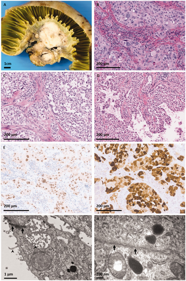
Figure 1.
Patient #1, presenting with a peritoneal mass. A, Grossly, the resected tumor was a solid tanwhite mass centered in the small bowel mesentery, abutting the muscularis propria but not invading into the superficial bowel tissue layers. On hematoxylin and eosin stained microscopic sections, the tumor was predominantly composed of nests or sheets of epithelioid cells with round vesicular nuclei of varying diameters and (B) eosinophilic or (C) clear cytoplasm. D, Papillary structures were seen in a minority of sections. Immunohistochemistry for (E) WT-1 and (F) calretinin was positive in tumor cells. Electron microscopy revealed a poorly differentiated tumor with (G) microvilli (arrowhead) and desmosomes (arrow). H, There was evidence of basement membrane formation (arrowhead).

