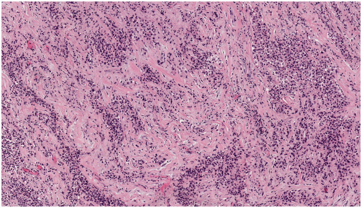
Figure 2.
Patient #2, presenting with a pericardial mass. On hematoxylin and eosin stained microscopic sections, the tumor showed infiltrating cords and nests of epithelioid cells within a sclerotic stroma. The cells were monomorphic with hyperchromatic eccentric nuclei and eosinophilic cytoplasm. Photomicrograph taken at 100× original magnification.

