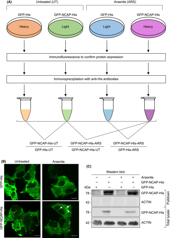
Fig. 1.

Identification of NCAP‐interacting proteins. (A) Scheme depicting the identification of NCAP‐interacting proteins by immunoprecipitation and SILAC mass spectrometry. (B) Localization of GFP‐NCAP‐His in A549 cells. Note that NCAP is mainly present as diffused in the untreated cells (red arrows) while it is present as both aggregated (white arrows) and diffused (red arrows) in the arsenite‐treated cells. A549 cells transfected with GFP‐His is used as the control. Scale, 10 μm. (C) western blot showing the pulldown of GFP‐NCAP‐His from A549 cells untreated or treated with arsenite. GFP‐His is used as a control in the transfection.
