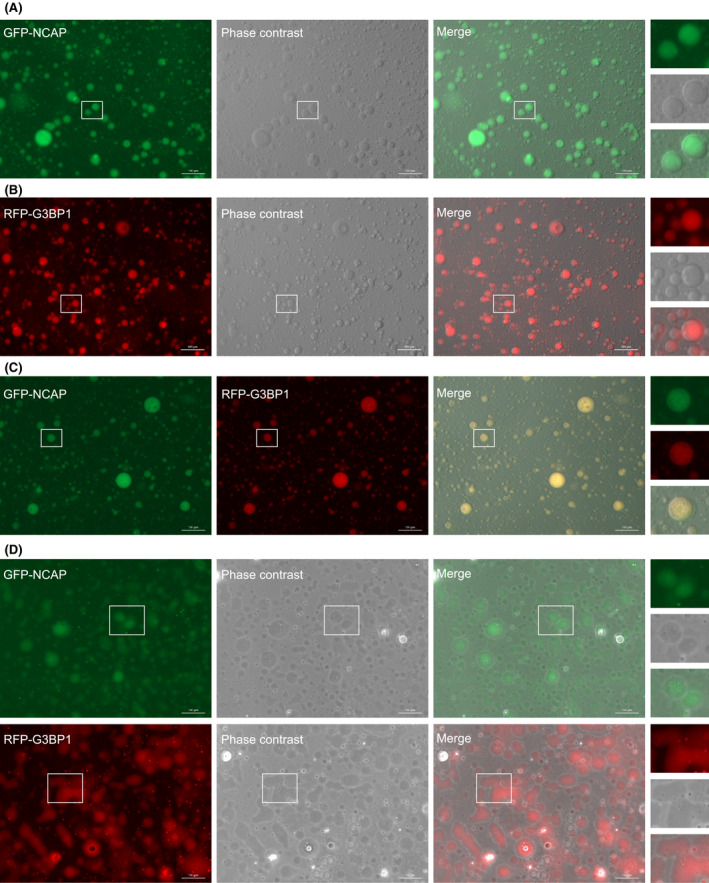
Fig. 6.

NCAP undergoes LLPS to form liquid droplets. (A) GFP‐NCAP is mixed with RNA to induce phase separation. The images were captured in a fluorescence microscope under 20× objective. (B) RFP‐G3BP1 is similarly phase separated with RNA. (C) GFP‐NCAP and RFP‐G3BP1 droplets were mixed and imaged. Note that droplets formed by both GFP‐NCAP and RFP‐G3BP1 colocalize. (D) GFP‐NCAP or RFP‐G3BP1 has liquid‐like properties. Droplets formed by GFP‐NCAP or RFP‐G3BP1 were allowed to stay on the surface for > 20 min and imaged. Note that the droplets flattened with time and wetted the surface showing their liquid property. A part of the image is enlarged and displayed on the right side of respective panels. Scale, 100 μm.
