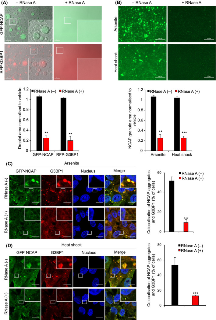
Fig. 7.

Phase separation and SG association of NCAP requires RNA. (A) GFP‐NCAP or RFP‐G3BP1 droplets phase separated with RNA were incubated in presence or absence of RNase A. Note that RNase A treatment dissolved both GFP‐NCAP and RFP‐G3BP1 droplets. Scale, 100 μm. The bottom panel shows the quantification of phase separation. (B) GFP‐NCAP expressing cells were treated with 100 μm arsenite or heat‐shocked at 41 °C for 1 h. GFP‐NCAP granules were isolated from the cells and incubated with or without RNase A for 6 h. The samples were observed under a fluorescence microscope. Note that arsenite treatment‐ or heat‐shock‐induced granules were significantly dissolved in the presence of RNase A, confirming the requirement of RNA for the integrity of NCAP granules. Scale, 100 μm. The bottom panel shows the quantification. (C‐D) GFP‐NCAP expressing A549 cells were untreated or treated with 100 μm arsenite (C) or heat‐shocked at 41 °C (D) for 50 min. The cells were gently permeabilized with detergent, and the live cells were untreated or treated with RNase A for 10 min. The fixed cells were subjected to IF using anti‐G3BP1 antibodies. Note that treatment with RNase A significantly reduced the accumulation of GFP‐NCAP in SGs. Scale, 10 μm. The right‐side panels show quantification of GFP‐NCAP association with SGs. Mean values ± SEM are shown for three independent experiments; **P < 0.01, ***P < 0.001.
