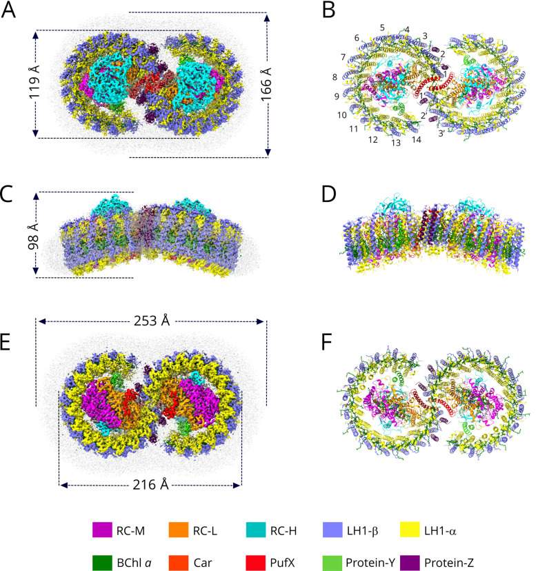
Figure 1. Cryo-EM structure of the dimeric RC-LH1 complex from Rba. sphaeroides.
(A,C,E) Views of the density map, coloured as in the key at the bottom of the figure. Detergent and other disordered molecules are in grey. (B,E,F) Ribbon models of the complex, made using ChimeraX [34]. (A) View of the cytoplasmic face of the density map of the complex, showing the diameters of the short axes of the detergent belt and the complex. (B) View as in (A), as a ribbon model; the LH1 subunits are numbered. (C) View of the density map in the plane of the membrane showing the height of the complex. (D) View as in (C), as a ribbon model. (E) Perpendicular view of the density map from the periplasmic side with measurements of the long-axis of the complex and detergent micelle. (F) Ribbon model corresponding to (E).

