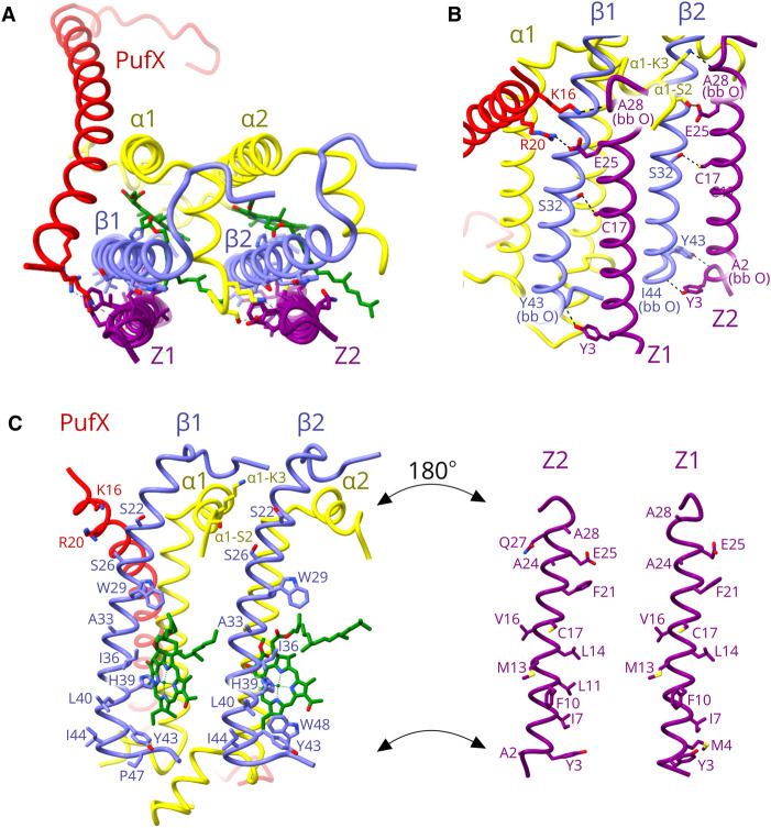
Figure 2. The positions and transmembrane interactions of the two protein-Z polypeptides.
(A) View of part of the LH1 ring near the monomer–monomer interface, perpendicular to the membrane from the cytoplasmic side. The transmembrane regions of protein-Z1 and protein-Z2 are closely appressed against the LH1 β1 and β2 polypeptides. (B) View in the plane of the membrane, showing hydrogen bonds (dashed lines) between Z1/Z2 and the respective β1 and β2 polypeptides. Other bonds with LH1 α1 and PufX near the cytoplasmic face of the membrane are labelled. Supplementary Table S2 lists the hydrogen bonds relating to Figure 2B. (C) ‘Open book’ format to show the opposing, interacting hydrophobic faces of Z1 and Z2 and the respective β1 and β2 polypeptides. The labelled residues are predicted to be in van der Waals contact.

