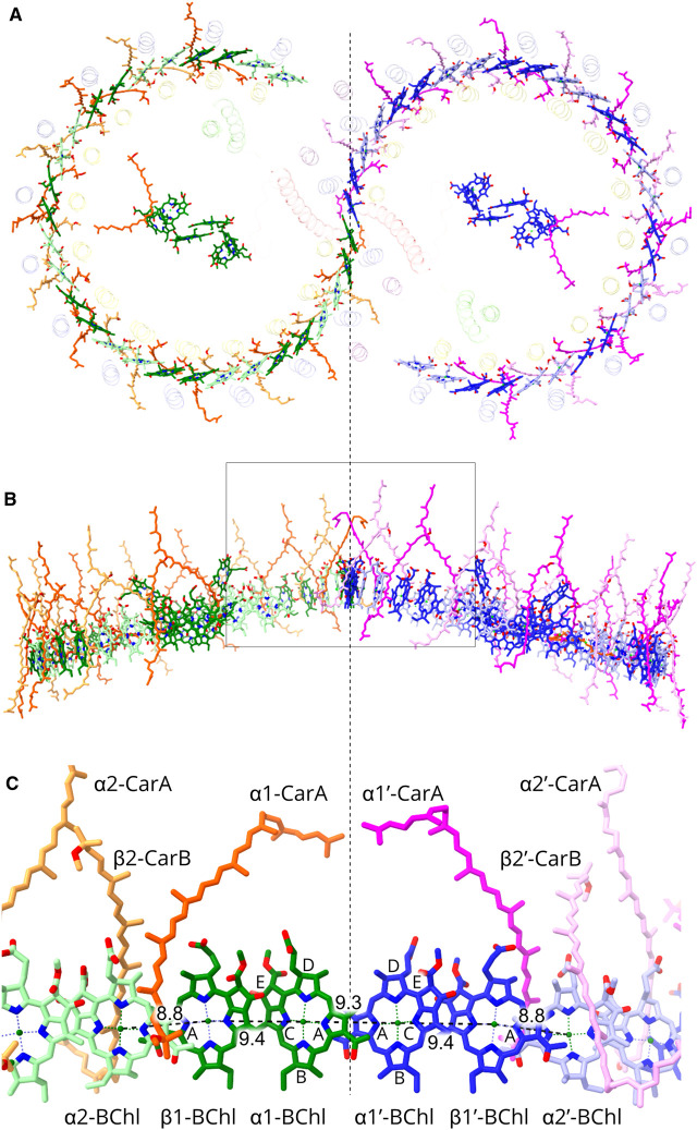
Figure 6. The bacteriochlorophyll and carotenoid pigments in the RC-LH1 dimer.
Proteins are faded for clarity. The dashed line divides the complex into the two halves. The central pigments belong to the RCs. (A) View from the periplasmic side of the membrane showing the two arcs of 28 BChls. Within one monomer half the BChls are coloured in two shades of green to distinguish between pairs belonging to individual LH1 αβ subunits, and in the other half two shades of blue are used. Similarly, two shades of orange or magenta indicate pairs of carotenoids belonging to individual LH1 αβ subunits on each half of the dimer. (B) View in the plane of the membrane. The box indicates the interface zone magnified in panel (C). (C) BChl and carotenoid pigments at the interface, coloured as in (A), and with the α and β polypeptides numbered as in Figure 1B. Rings A–E of the BChl macrocycles are labelled, and Mg–Mg distances are shown in Ångstroms for intra- and inter-subunit BChls.

