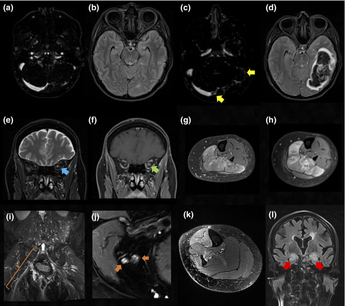
FIGURE 2.

Radiological findings in patients with autoimmunity following SARS‐CoV‐2 vaccinations. (a), (b) Four days prior to the onset of supraventricular tachycardia symptoms: aside from a hypoplastic left sigmoid sinus findings are unremarkable in contrast‐enhanced magnetic resonance venography (CE‐MRV) (a) and axial fluid attenuated inversion recovery (FLAIR) sequence (b). (c) Follow‐up CE‐MRV reveals an occlusive thrombus of the left sigmoid sinus and a non‐occlusive thrombus of the right transverse sinus (yellow arrows). (d) The corresponding axial FLAIR sequence shows resulting congestive bleeding in the left temporal and occipital lobe. (e) Coronal T2‐weighted (T2w) and (f) gadolinium contrast‐enhanced (Gd CE) T1w orbital sequences reveal T2w hyperintensity (e) (blue arrow) and associated mild contrast enhancement of the left optic nerve (f) (green arrow) consistent with optic neuritis. (g), (h) Axial, fat saturated, Gd CE T1w saturated images at the level of the left lower leg 6 weeks before (g) and 7 days after (h) vaccination demonstrate progressive contrast uptake of the left soleus muscle indicating reactivation of focal myositis. (i) Coronal T2w MRI reveals hyperintense enlargement of the right L5 nerve root (orange bracket) consistent with L5 radiculitis. (j), (k) Axial T2w sequence with spectral fat saturation at the level of the distal right thigh (j) and right lower leg (k) are shown. Somatotopic L5 lesion pattern with hyperintense, fascicular enlargement of the common peroneal nerve and the ventral section of the tibial nerve (j) (orange arrows) and subsequent denervation oedema of the fibularis longus, extensor digitorum longus, tibialis anterior and posterior muscles (k) are evident. (l) Coronal FLAIR sequence demonstrates bilateral hippocampal hyperintensities (red arrows) and mild swelling in line with limbic encephalitis [Color figure can be viewed at wileyonlinelibrary.com]
