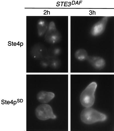
FIG. 3.
Localization of GFP-Ste4p and GFP-Ste4pSD10 in STE3DAF cells. Strain AC17-2B (MATa STE3DAF ste4::HIS3) containing a GFP-STE4 plasmid (pBTL49) or a GFP-STE4SD10 plasmid (YCpGFP-SD10) was treated with α-factor (0.1 μM) for the indicated periods of time. The live cells were viewed by fluorescence microscopy using an FITC filter set.

