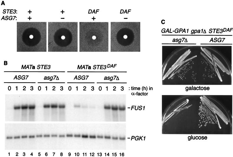
FIG. 4.
Effect of ASG7 on signaling in STE3DAF cells. (A) Halo assays were performed with 5 μl of 1 mM α-factor using the following strains (from left to right): H67-9D.Ba, a MATa STE3 ASG7 strain, H67.9D.a7, a MATa STE3 asg7::URA3 strain, H67-6C.Ba, a MATa STE3DAF ASG7 strain, and H67-6C.a7, a MATa STE3DAF asg7::URA3 strain. (B) The following strains were treated with α-factor (0.1 μM) for the indicated periods of time: H67-9D.Ba, a MATa STE3 ASG7 strain (lanes 1 to 4), H67-9D.a7, a MATa STE3 asg7::URA3 strain (lanes 5 to 8), H67-6C.Ba, a MATa STE3DAF ASG7 strain (lanes 9 to 12), and H67-6C.a7, a MATa STE3DAF asg7::URA3 strain (lanes 13 to 16). RNA was isolated, transferred to nitrocellulose, hybridized with a FUS1 probe, and rehybridized with a PGK1 probe. (C) MATa STE3DAF gpa1::TRP1 ASG7 (H125-7D) and MATa STE3DAF gpa1::TRP1 asg7::HIS3 (H125-7D.aH) strains containing a plasmid with GPA1 under the control of the GAL promoter were streaked onto galactose- or glucose-containing plates and grown at 30°C for 2 to 3 days.

