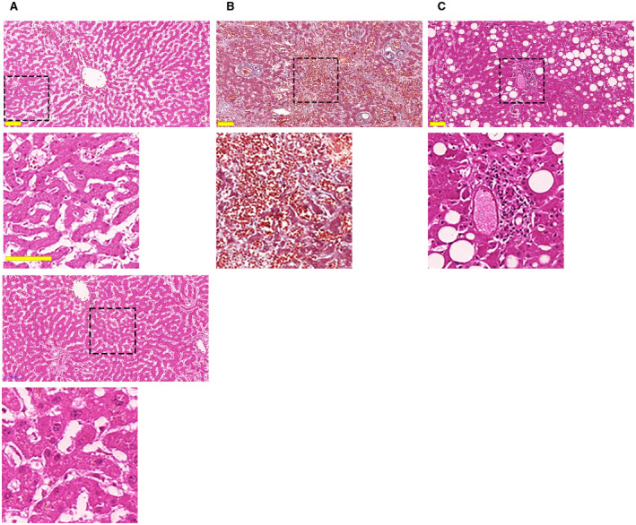
FIG. 1.

COVID‐19 infection is associated with ischemic necrosis, congestion, steatosis, and portal inflammation in most patients. (A) Representative hematoxylin and eosin staining of a liver section demonstrates areas of necrosis as evidenced by the uniform cytosolic staining and absence of nuclei. This pattern was observed in 26 of 27 patients. A magnified view of the dotted square area is shown in the inset below. The bottom panel and inset show an area not affected by necrosis, as demonstrated by the light eosin staining and presence of normal‐sized nuclei. (B) Masson’s trichrome staining shows diffuse blood retention and congestion in liver sinusoids (inset). This was observed in 23 of 27 patients. (C) Areas of macrovesicular steatosis, as highlighted by the typical round negative images, in the liver of a representative patient with COVID‐19. This was observed in 17 of 27 patients. Representative area of portal inflammation, as shown by the presence of inflammatory infiltrate surrounding portal tracts, were similarly observed (inset) in 14 of 27 patients. Scale bar, 100 μm.
