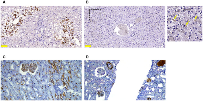
FIG. 2.

Expression of activated coagulation factors is uncommon in COVID‐19 liver specimens. (A) Positive C4d immunohistochemical staining (brown), a marker of activated complement cascade, was observed in 22% of the liver samples from patients with COVID‐19. (B) Positive staining for CD61 was detected in 14% of the 27 COVID‐19 liver samples. Light brown staining (arrows in the inset) is seen along the sinusoidal walls. Scale bar, 100 μm. Positive CD61 was present in control samples of kidney tissue (data not shown). (C) Positive control staining for C4d in kidney tissue from a transplant rejection allograft. (D) Cd5 positive staining was similarly found in kidney tissue from the same patient.
