Figure 2.

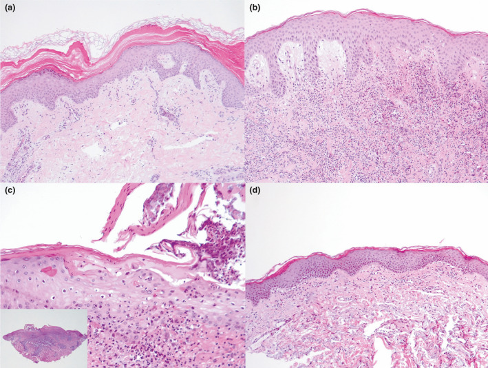
(a–e) Histological analysis of incisional cutaneous biopsy taken from the patients: (a) pityriasis rubra pilaris, showing acanthotic epidermis with fusion and broadening of rete ridges and alternating orthokeratosis and parakeratosis; (b) mildly acanthotic epidermis with subepidermal oedema and intense interstitial neutrophilic infiltrate in the dermal layer in a patient with Sweet syndrome; (c) pityriasis lichenoides et varioliformis acuta, showing focal epidermal ulceration, spongiosis, parakeratosis, and interface inflammation within a wedge‐shaped dermal inflammatory cell infiltrate; and (d) vacuolar interface dermatitis with a dermal inflammatory lymphohistiocytic infiltrate and a mild epidermal spongiosis in a patient with erythema multiforme. Haematoxylin and eosin, original magnification (a) × 200; (b,c) × 50; (c) × 400, inset × 25.
