
Figure 2.
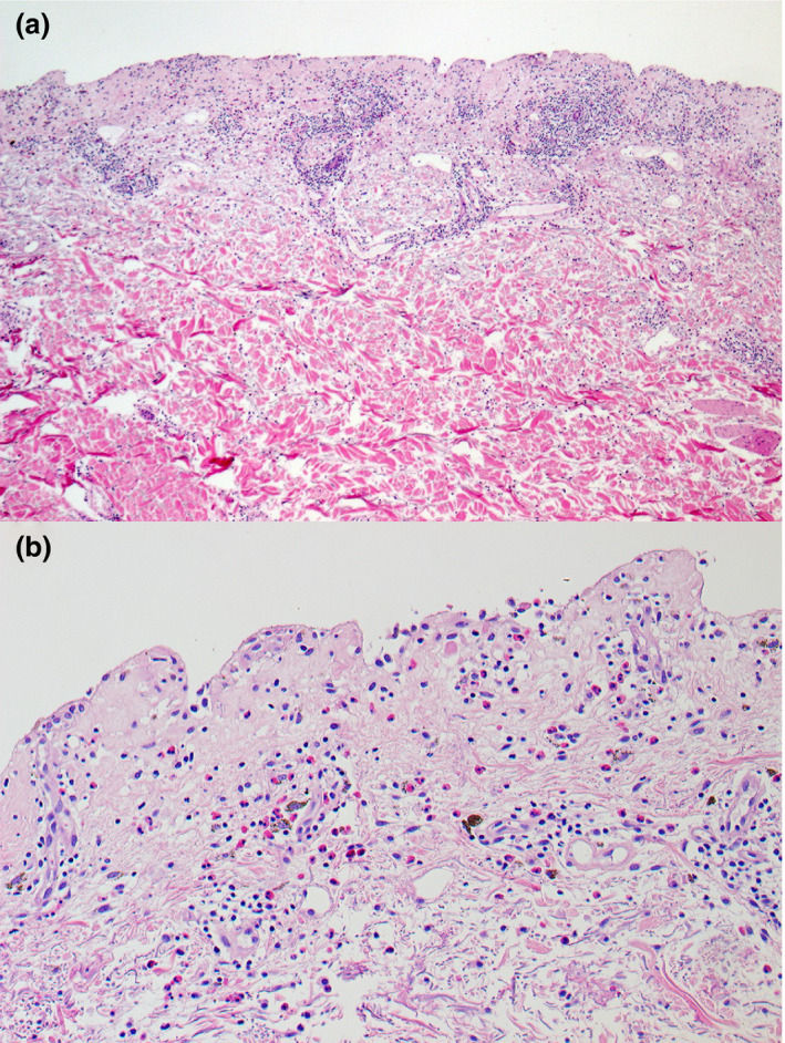
(a,b) Histological examination of a punch biopsy was performed from the lesion on the patient's back showed (a) subepidermal separation with superficial and deep perivascular inflammatory cell infiltration and (b) mixed inflammatory cells infiltrate, composing of lymphohistiocytes and numerous eosinophils. Melanophages were seen in the upper dermis. Haematoxylin and eosin, original magnification (a) × 50; (b) × 200.