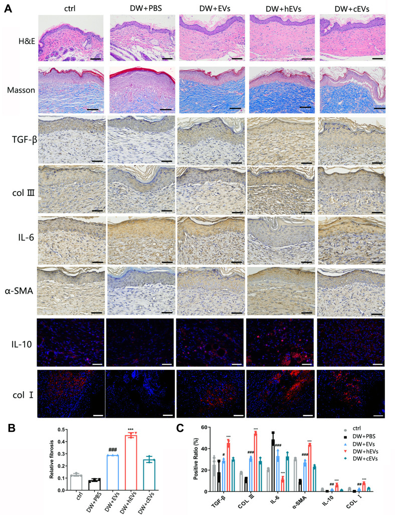
Figure 7.
Observation of wound healing quality and velocity after operation. (A) HE and Masson staining of tissue wounds 14 days after operation and expression of IL-6 and IL-10 in wound tissue was observed 7 days after operation. Expression of α-SMA, TGF-β, COLI, COLIII in wound tissue was observed 21 days after operation. n=3. (B and C) Masson, immunofluorescence and immunohistochemical statistical analysis. Data are represented as mean ± SD. *, vs DW+EVs; #, vs DW+PBS; n = 6. **P < 0.001, ***P < 0.0001, #P < 0.05, ##P < 0.001, ###P < 0.0001. Bars, 100μm.

