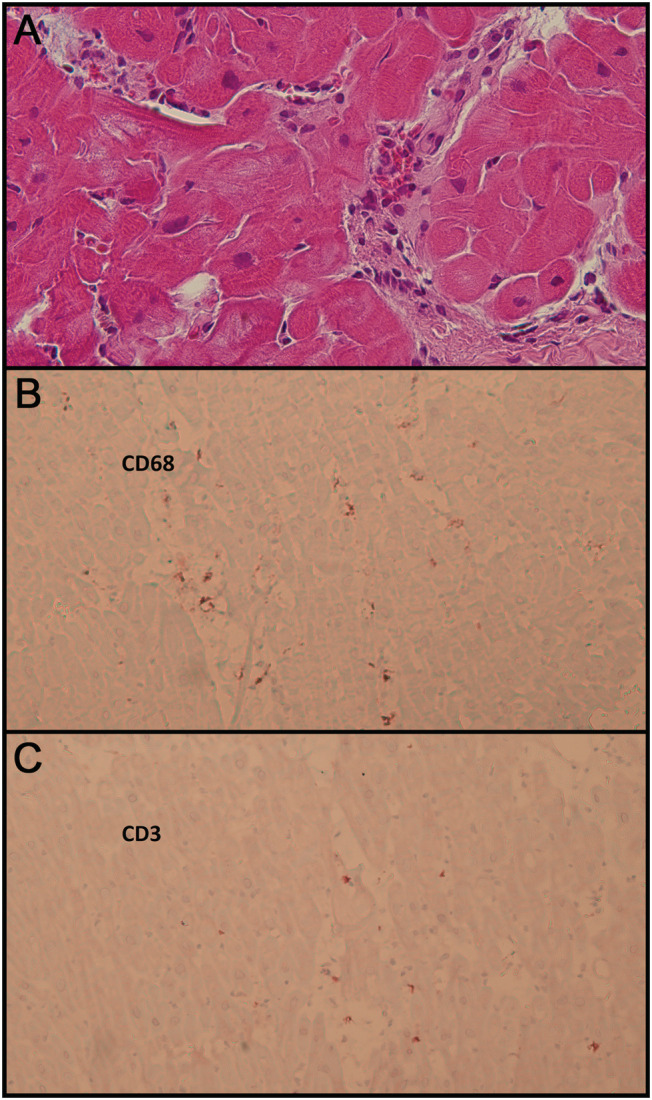
Figure 2.

Histology and immunohistology of endomyocardial biopsy. (A) Haematoxylin and eosin stain, primary magnification 40:1. Infiltration of mononuclear inflammatory cells (macrophages and lymphocytes) with mild interstitial oedema. Non‐hypertrophic myofibres with regular nuclei and cytoplasm without degeneration or necrosis. (B) Immunohistochemistry, CD68, anti‐alkaline phosphatase method, primary magnification 20:1. Considerably increased number of macrophages. (C) Immunohistochemistry, CD3, anti‐alkaline phosphatase method, primary magnification 20:1. Moderately increased number of T lymphocytes.
