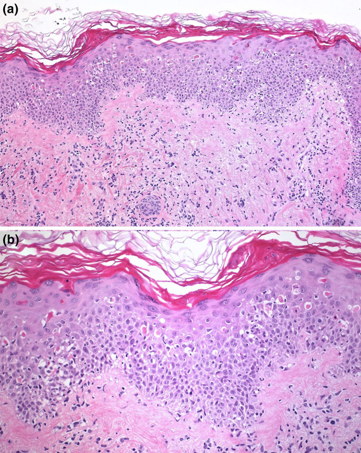
Figure 2.

(a) Skin biopsy demonstrated moderate perivascular, dermal lymphoid infiltration with focal interface dermatitis (Hematoxylin‐eosin, 100×). (b) Higher magnification revealed multiple apoptotic keratinocytes at all epidermal layers, sometimes with satellite lymphocytes (Hematoxylin‐eosin, 200×)
