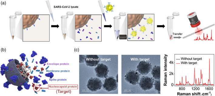
FIGURE 5.

(a) Schematic illustration for the surface‐enhanced Raman scattering‐based immunoassay of severe acute respiratory syndrome coronavirus 2 (SARS‐CoV‐2) lysate using magnetic beads. (b) The structure of SARS‐CoV‐2 lysate containing various biological components. (c) Transmission electron microscopy images of magnetic immunocomplexes in the absence (left) and presence (right) of 102 PFU/ml of SARS‐CoV‐2‐lysate and the corresponding Raman spectra
