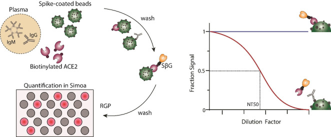
Figure 1.

Simoa neutralization assay. The assay is based on antibody‐mediated blockage of the ACE2‐spike interaction. Spike‐coated beads, biotinylated ACE2, and patient plasma are mixed. After several washing steps, streptavidin conjugated beta‐galactosidase (SβG) is added. The beads are then washed again, resuspended in resorufin β‐D‐galactopyranoside (RGP), and loaded into a microwell array for imaging. As the concentration of anti‐spike antibodies in the sample increases, the signal in the assay decreases. To determine neutralization capacity (NT50), the plasma sample is diluted and the signal at each dilution factor is normalized to the AEB of the highest dilution factor.
