FIGURE 2.

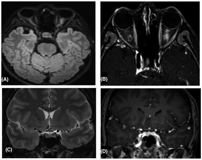
(A,B) Axial FLAIR and T1‐Gd weighted MRI evidencing swelling of the left optic nerve, enhancing after gadolinium administration. (C,D) Sagittal T2 and T1‐Gd weighted MRI, no enhancing of the optic chiasma is observed.

(A,B) Axial FLAIR and T1‐Gd weighted MRI evidencing swelling of the left optic nerve, enhancing after gadolinium administration. (C,D) Sagittal T2 and T1‐Gd weighted MRI, no enhancing of the optic chiasma is observed.