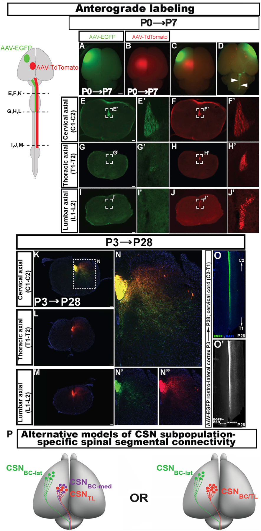
Figure 2. Early projection targeting specificity by CSN in the lateral cortex is durably maintained.

(A–D) Whole-mount view of a P7 brain injected at P0 with AAV-EGFP rostrolaterally (green, A) and AAV-tdTomato caudomedially (red, B; merged dorsal view showing both sites in C) to anterogradely label the CST (arrowheads, ventral view in D).
(E–J) Axial spinal sections from the same mouse at cervical (E and F), thoracic (G, H), and lumbar (I and J) segments. Only tdTomato+ axons extend to the lumbar cord. (E’–J’) Magnified views of the dorsal funiculus (boxed regions in EJ).
(K–M) Axial spinal sections from a P28 mouse that was similarly injected with the two AAVs at P3, at cervical (K), thoracic (L), and lumbar (M) levels. Only tdTomato+ axons extend to the lumbar cord.
(N–N’’) Magnified view of the boxed region in K. At P28, EGFP+ CSNBC-lat axons (green) extend collaterals into more ventral laminae in cervical gray matter compared to tdTomato+ CSNmedial axons (red) (individual channels in N’ and N’’).
(O and O’) Horizontal section of a cervical cord from the same mouse as in K–N (O). CSNBC-lat axons traverse and extend collaterals throughout the rostrocaudal extent of the cervical cord (O’).
(P) Schematic representation of two distinct possibilities that might theoretically give rise to segmentally distinct CSNmedial axon projections. In both, CSN in the lateral cortex (CSNBC-lat; green) project exclusively to the brainstem and cervical cord. (Left) CSNBC-med (purple) and CSNTL (red) are developmentally distinct, and this specification persists into maturity. (Right) CSNBC and CSNTL in the medial cortex are developmentally equivalent early, but later give rise to segmentally specific projections (CSNBC/TL in red).
Scale bars, 1 mm in A–D and 100 μm in E–O.
