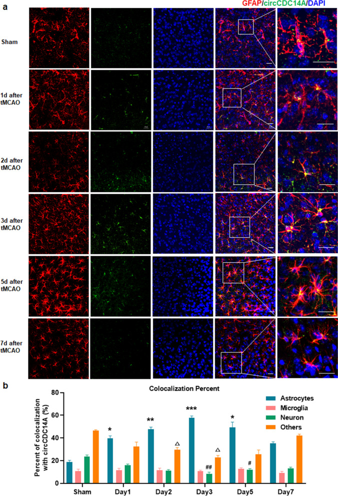
Fig. 2.
Colocalization analysis of circCDC14A with astrocytes, microglia, neuron and others in peri-infarct cortex. a Colocalization with GFAP in sham-operated and tMCAO mice from 1 to 7 days after modeling. Bar: 20 μm. b Quantification of circCDC14A colocalization percent with astrocytes, microglia, neuron and others, n = 3 mice per group. *P < 0.05, **P < 0.01, ***P < 0.001, compared to astrocytes in sham-operated mice; #P < 0.05, ##P < 0.01, compared to neuron in sham-operated mice; ∆P < 0.05, compared to others in sham-operated mice; two-way ANOVA test

