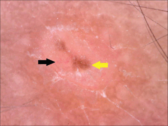
Figure 2.

Dermoscopy of papules on back shows follicular as well as extrafollicular lesions with central cluster of brown clods (yellow arrow) along with halo of pallor with telangiectasias (black arrow) and rim of fine white scales (170× magnification, Polarized mode)
