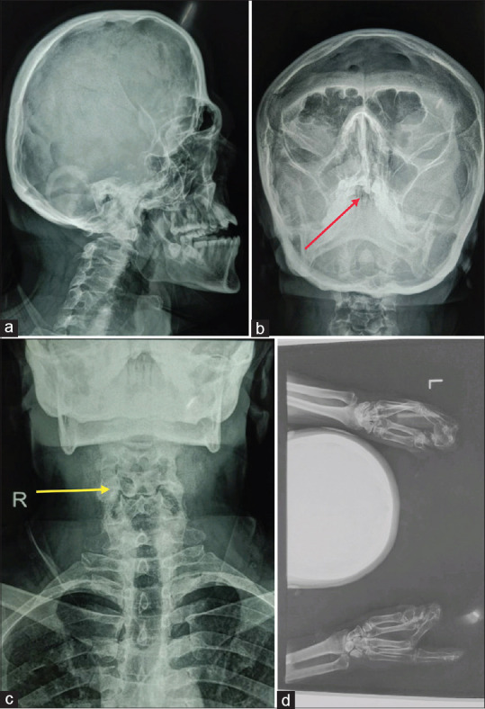
Figure 5.

(a) X-ray skull lateral view showing brachycephaly, hypoplastic maxilla, protrusion of mandible and small orbital cavity. (b) X-ray skull paranasal sinus showing cleft palate (Red arrow). (c) X-ray cervical spine showing blocked vertebrae at C5-C6 and C6-C7 level (yellow arrow). (d) X-ray bilateral hands showing complete syndactyly of both hands
