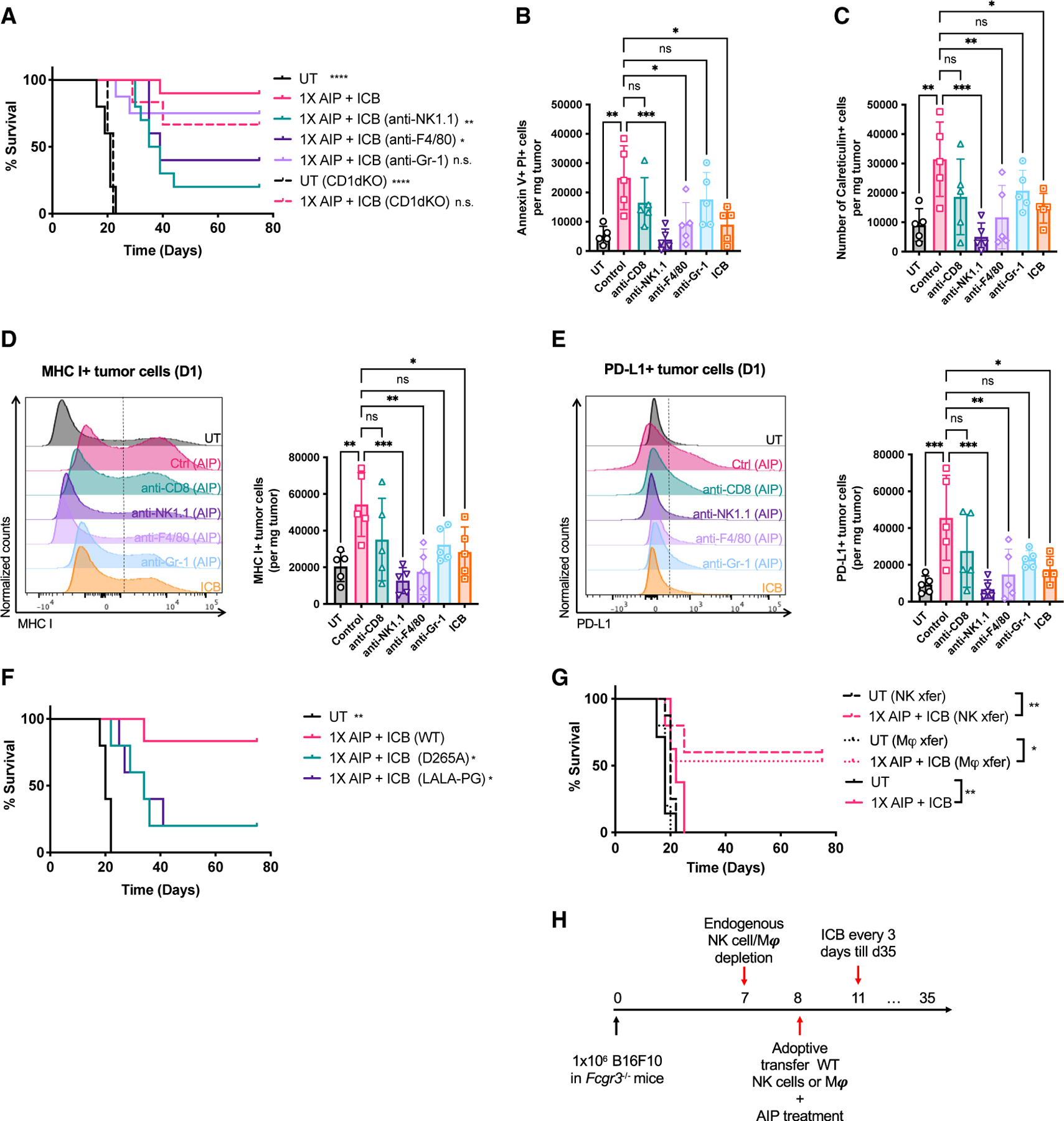
Figure 3. NK cells and macrophages play an important role in initial killing of tumor cells and tumor antigen delivery to DCs.

(A) Survival of B16F10 tumor-bearing mice over time with depleting antibodies for NK cells and macrophages injected 1 day before 1X AIP treatment (n = 5 mice/group, from one representative of two independent experiments).
(B and C) Quantification of apoptotic (B; Annexin V+, PI+) and surface calreticulin+ (C) tumor cells in UT, AIP-treated tumors with depleting antibodies for CD8+ T cells, NK cells, and macrophages 1 day before treatment.
(D and E) Representative flow cytometry histograms and quantification of MHC class I (D) and PD-L1 (E) expression on tumor cells (defined as live, CD45− and TA99+ cells) from mice 1 day post treatment as in (B) and (C).
(F) Survival over time following 1X AIP + ICB treatment in mice bearing B16F10 tumors (n = 5 mice/group), where D265A or LALA-PG mutations were introduced to the Fc portion of 2.5F-Fc.
(G and H) FcγRIII-deficient mice (n = 8–10 animals/group, pooled from two independent experiments) bearing B16F10 tumors were depleted of endogenous NK cells or macrophages 1 day prior to treatment with 1X AIP + ICB. One group of mice received adoptive transfer of 1 × 10⁶ WT NK cells (dashed lines), and another group received 3–4 × 10⁶ WT macrophages (dotted lines) injected on day 0. Tumor-bearing FcγRIII-deficient mice treated with or without 1X AIP + ICB (with no depletion or adoptive transfer) were used as a control (solid lines) Shown is survival of these mice over time (G) and the experiment timeline (H).
*p < 0.05; **p < 0.01; ***p < 0.001; ****p < 0.0001 versus 1X AIP + ICB by log rank test in (A), (F), and (G) and by one-way ANOVA followed by Dunnett post test in (B) and (C) and (D) and (E). All error bars are SD. See also Figure S3.
