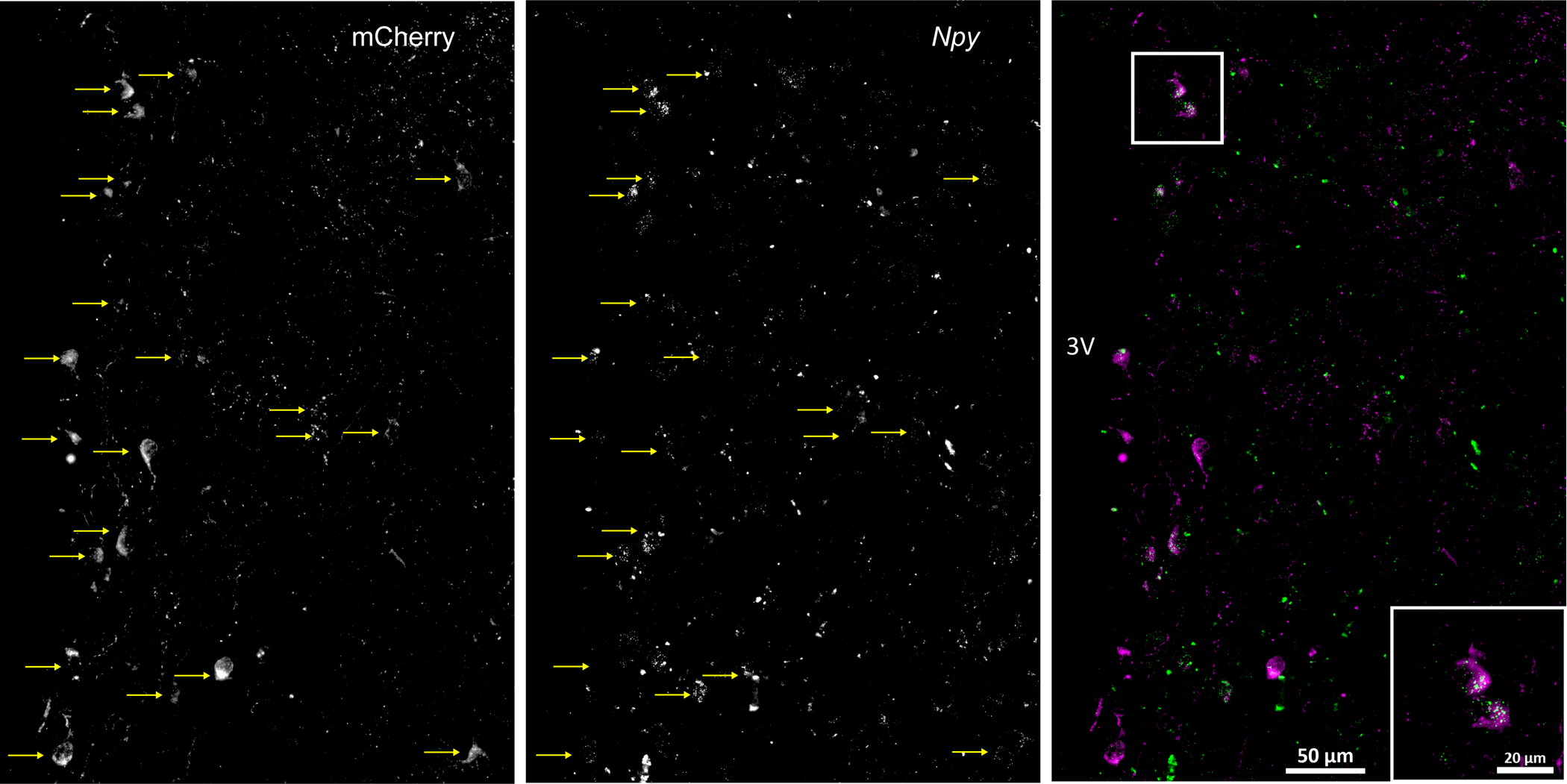
Fig. 1. Selective expression of AAV-Syn-mCherry in neurons that express Npy1r.

Male npy1r-cre mice received unilateral PVN injections of a cre-dependent adeno-associated virus (AAV), allowing for the selective expression of a fluorescent protein (mCherry; left panel) in synaptosomes, which can be visualized in the cell bodies, axon fibers, and synapses of NPY Y1R-containing neurons (middle panel). Images show that 95% (19/20) of neurons that expressed mCherry (magenta) also expressed mRNA for the NPY1R (green puncta; at least 3 per neuron), quantified using RNAScope (right panel).
