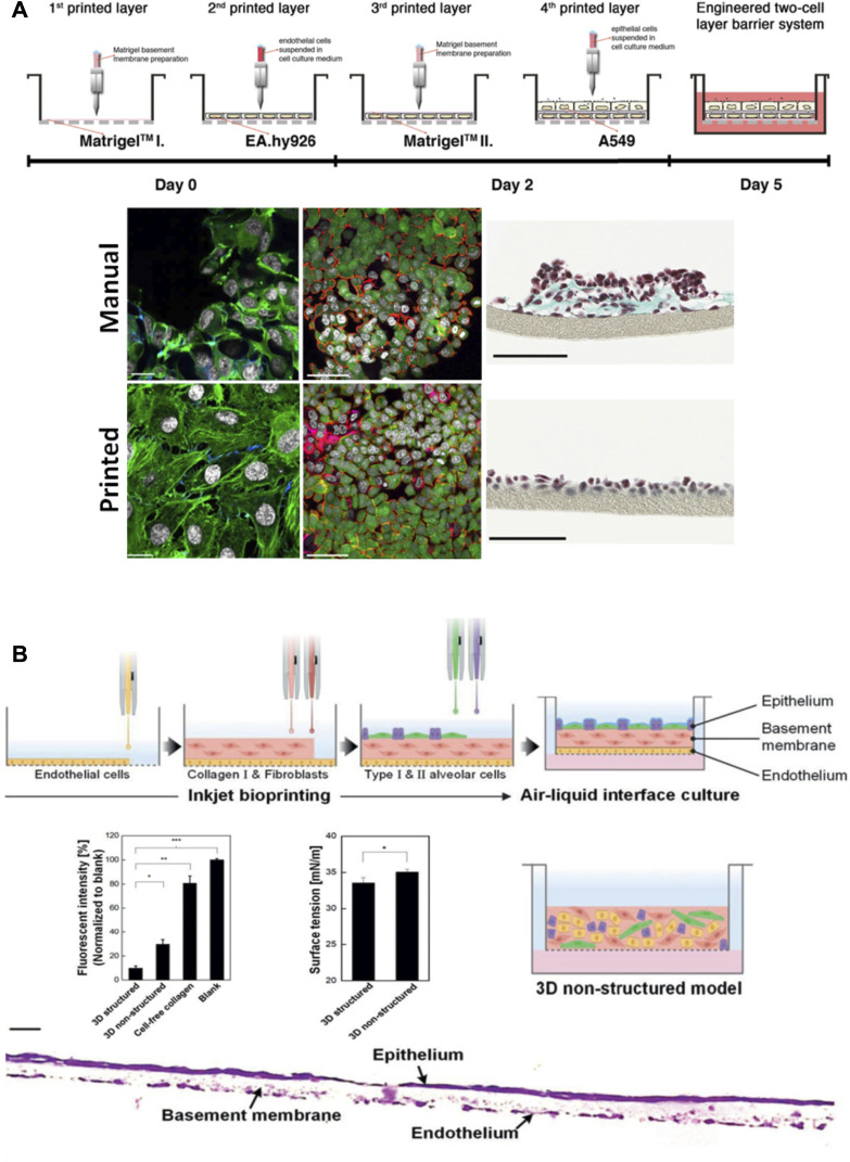
FIGURE 8.
Models that recreate the air-blood barrier by using 3D printing approaches. (A) Schematic representation of the process to fabricate an air-blood tissue barrier analog composed of endothelial cell, basement membrane and epithelial cell layers. Comparison of the final cell distribution when seeding cells manually versus bioprinting them: immunofluorescence labelling of F-actin cytoskeleton (green) and nuclei (grey) (scale bar 20 µm), laser scanning micrographs (scale bar 50 µm) and brightfield micrographs (scale bars 100 µm). Reproduced from (Horvath et al., 2015). (B) Schematic representation of the fabrication process of an alveolar barrier model composed of alveolar cells, lung fibroblasts, and lung microvascular endothelial cells. Comparison of the 3D model and the 3D unstructured model used as control, and a cross sectional image of the stained barrier (scale bar 20 µm). Reproduced from (Kang et al., 2021).

