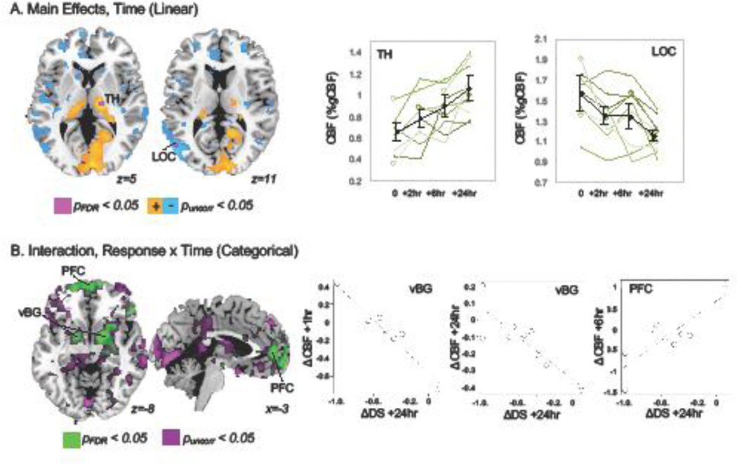
Figure 1.
A. Monotonic changes in CBF were noted after ketamine infusion in patients with depression. A cluster in the thalamus (TH) exhibited significant increased CBF over time, while a cluster in lateral occipital cortex (LOC) showed significant decreases, pFDR < 0.05 (green). At more permissive thresholds (puncorr < 0.05, orange/blue), decreased CBF was also noted in anterior cingulate and dorsomedial prefrontal cortex, and increased CBF was noted in medial visual cortex. Mean CBF (% global CBF) is plotted for TH and LOC clusters in the top-right panels for all patients in black and for each individual subject in shades of green to demonstrate consistency of these changes across subjects. Subjects with missing data are plotted with open circles. B. Non-monotonic changes in CBF differed according to antidepressant response in time-by-response interactions. Bilateral ventral basal ganglia (vBG, including pallidum and ventral striatum) and medial prefrontal regions (PFC) exhibited significant interactions, pFDR <0.05 (green). Scatter plots display relationships between change in (delta, Δ) in CBF and change in depression symptoms (averaged across three inventories) meeting statistical criterion in post hoc analyses (pBonf < 0.05).

