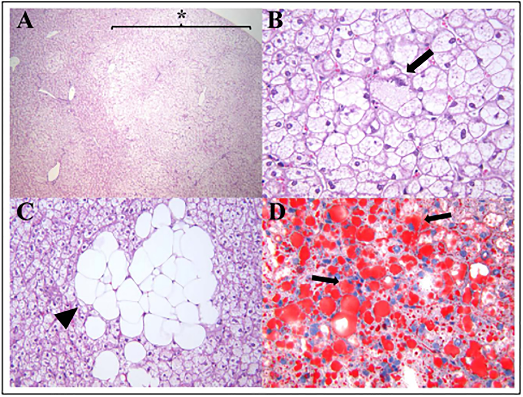
Figure 1.

Histological changes associated with hepatic steatosis in a 14-year-old female marmoset. (A) Sections of liver have multifocal large regions of microvesicular steatosis extending from periacinar to periportal regions (star, 40x). (B) Areas of microvesicular steatosis are characterized by swollen hepatocytes containing small clear intracytoplasmic vacuoles and eccentrically displaced nuclei (arrow, 400x). (C) Higher magnification of macrovesicular steatosis demonstrating clusters of hepatocytes with large intracytoplasmic lipid vacuoles completely effacing the nuclei (arrowhead; 400x). (D) Lipid content in areas of microvesicular and macrovesicular hepatic steatosis is demonstrated by Oil red-O staining of hepatocytes (red, arrows; 400x).
