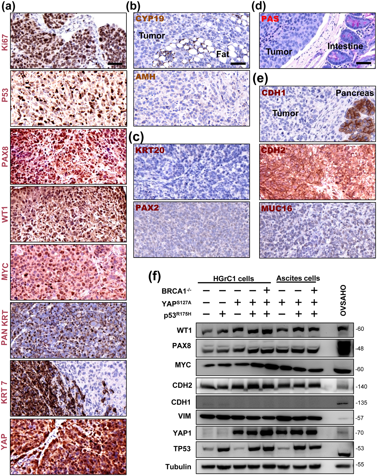
Figure 6. YAP1 induced High grade ovarian cancers with serous features from less-differentiated granulosa cells.

(a) Molecular features of tumors derived from the xenograft mouse model of YAPS127A/TP53R175H/BRCA1−/− cells. Representative image showing high level expression of Ki-67, nuclear TP53, PAN keratin (PAN KRT), Keratin 7 (KRT7), PAX8, WT-1, MYC, and YAP1 protein in tumor tissues analyzed by immunohistochemistry. Scale bar: 50 μm. (b) Representative images showing that tumor tissues are negative for the expression of granulosa cell tumor markers (CYP19A1 and AMH). (c) Representative images showing that tumor tissues are negative for low grade serous carcinoma and mucinous ovarian cancer markers (PAX2 and KRT20). (d) Representative images showing that tumor tissues are negative for mucinous cancer markers (PAS staining). (e) Representative images showing that tumor tissues have high expression of CDH2 (N-cadherin), but low expression of CDH1 (E-cadherin) and MUC16 (CA125), which is a molecular feature of the recently identified mesenchymal type of ovarian high grade serous cancer. Scale bar: 50 μm. (f) Representative blots showing expression of WT1, PAX8, MYC, CDH2 (N-cadherin), CDH1 (E-cadherin), VIM (vimentin), TP53, and in established HGrC1-CTL cells, HGrC1- TP53R175H cells, HGrC1-YAPS127A cells, HGrC1-YAPS127A/TP53R175H cells, and HGrC1-YAPS127A/TP53R175H/BRCA1−/− cells. Expressions of these biomarkers were also examined in ascites tumor cells collected from immunodeficient mice carrying tumors from HGrC1-YAPS127A, HGrC1-YAPS127A/TP53R175H, and HGrC1-YAPS127A/TP53R175H/BRCA1−/− cells. OVSAHO cells were used as control.
