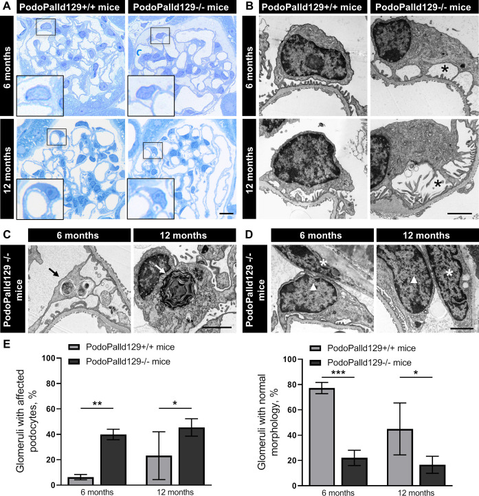
Fig 2. Podocytes of PodoPalld129-/- mice show morphological abnormalities.
(A) In Richardson’s stained semithin sections of PodoPalld129-/- mice we have found podocytes with enlarged subpodocyte space compared with controls as illustrated in the higher magnifications. Scale bar represents 10 μm. (B) TEM images show podocytes with enlarged subpodocyte space in both age groups of PodoPalld129-/- mice (asterisks). (C, D) Podocytes with autophagosomes/laminar bodies (arrows) and contacts between parietal epithelial cells (asterisks) and podocytes (arrowhead) were found in TEM images of both age groups in PodPalld129-/- mice. Scale bars represent 2 μm. (E) PodoPalld129-/- mice had significantly more glomeruli with affected podocytes and correspondingly significantly fewer glomeruli without abnormalities than controls of the same age (mean±SD, * p<0.05, ** p<0.01 and *** p<0.001; two-way ANOVA with FDR correction).

