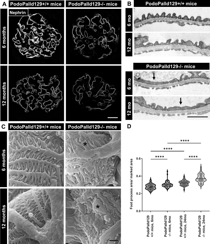
Fig 3. PodoPalld129-/- mice have effaced podocyte foot processes.
(A) Immunofluorescence staining of kidney sections shows a reduced expression of the slit membrane protein nephrin in PodoPalld129-/- mice compared with corresponding controls. Scale bar represents 20 μm. (B) Transmission electron micrographs (arrows) and (C) scanning electron micrographs (asterisks) revealed strongly effaced podocyte foot processes in PodoPalld129-/- mice. Scale bars represents 1 μm. (D) Quantification of foot process area confirmed the increased podocyte foot process effacement in 6 months as wells as 12 months old PodoPalld129-/- mice compared with corresponding controls. Data are represented as violin plot (**** p<0.0001; two-way ANOVA with FDR correction).

