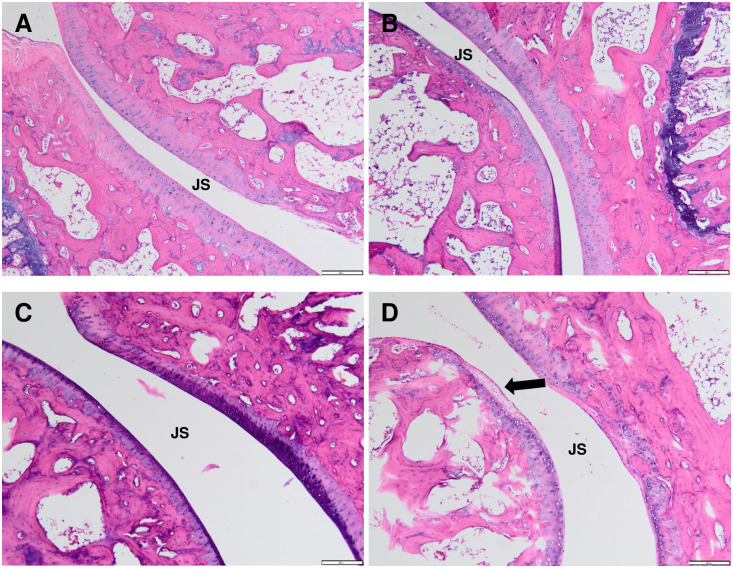
Fig 10. Histological sections of contralateral ankle joint by haematoxylin and eosin (H&E) staining at 40x magnification in control (A), polyarthritic rat inoculated with CFA at 5.0 mg/mL (CFA 5.0) (B), 7.5 mg/mL (CFA 7.5) (C) and 10.0 mg/mL (CFA 10.0) (D).
(A) Like the ipsilateral ankle joint, the control rat exhibited normal joint cartilage structure, regular joint space with the normal connective tissue of the synovial membrane. CFA 5.0 group did not demonstrate prominent inflammatory cells penetration in the regular joint space. Meanwhile, the inflammatory cells infiltration was identified between the joint space (arrow) of the CFA 7.5 (C) and CFA 10.0 (D) groups, although it was not as extensive as the ipsilateral ankle joint.

