
FIGURE 1.
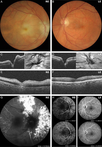
Unilateral optic nerve head swelling and retinal ischemia. Retinal photography of the right fundus demonstrated a swollen, pale optic nerve head, with generalized retinal ischemia evidenced by cotton wool spots and a “cherry-red spot” within the fovea (A). The left fundus by contrast was grossly normal (B). Optic nerve head spectral-domain optical coherence tomography (SD-OCT) imaging of the right eye demonstrated predominantly nasal optic nerve head swelling evident in horizontal cross-section and 3-dimensional projection (C). Left optic nerve head SD-OCT images were unremarkable (D). Horizontal cross-sectional SD-OCT imaging of the right fovea demonstrated hyperreflectivity of the inner retinal layers, characteristic of retinal ischemia (E). SD-OCT imaging of the left fovea, by comparison, was unremarkable (F). Representative fundus fluorescein angiography imaging of the right eye demonstrated profoundly delayed arterial perfusion with patchy choroidal perfusion at 0:55 minutes (G). Retinal arterial perfusion was first observed at 1:50 minutes, with venous and incomplete choroidal perfusion observed at 5:03 minutes (H). Fundus fluorescein angiography imaging of the left eye demonstrated normal arterial, venous, and choroidal perfusion at similar time points of 1:28 and 5:22 minutes following intravenous fluorescein bolus infusion (I). LE indicates left eye; RE, right eye.