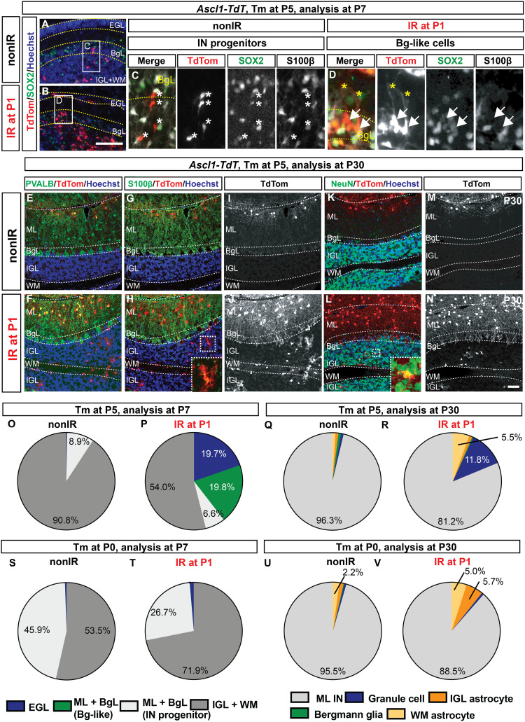
Fig. 5. Injury-induced Ascl1+ BgL-NEPs give rise to granule cells and astrocytes but not Bg.
(A and B) IF analysis of Ascl1-TdT brains at P7 (48 hours after Tm). (C and D) Insets show a migrating IN progenitors [asterisks; (C)] and a SOX2+ S100β− BgL-like Ascl1-TdT cell [arrows; (D)]. Yellow asterisks show the pial projection of the TdT+ cells. (E to N) IF analysis of nonIR and IR P30 Ascl1-TdT cerebellum that was given Tm at P5 showing examples of the TdT+ ML INs (PVALB), astrocytes (S100β), and granule cells (NeuN in the IGL). (O to V) Proportions of TdT+ cells at P7 (O. P, S, and T) or at P30 (Q, R, U, and V) from nonIR (O, Q, S, and U) and IR (P, R, T, and V) Ascl1-TdT cerebella that are given Tm at P5 [(O to R), n ≥ 3 brains per condition] or at P0 [(S to V) n = 3 brains per condition]. Only percent values that are ≥2 are shown in the graphs (refer to table S9 for all the values). Scale bars, 100 μm (B to G) and 50 μm (K to T).

