Figure 2.
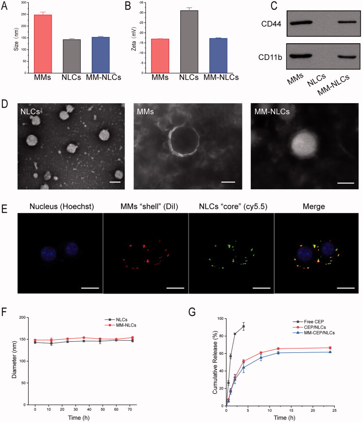
Characterization of the MM-coated biomimetic nanoparticles. (A) The size and (B) zeta potentials of MMs, NLCs and MM-NLCs (n = 3, mean ± SD). (C) Western blot results of CD44 and CD11b in MMs, NLCs and MM-NLCs. (D) TEM images of NLCs, MMs, and MM-NLCs (scale bar = 100 nm). (E) CLSM images of the colocalization of the nucleus (blue), MMs ‘shell’ (green) and NLCs ‘core’ (red) (scale bar = 10 μm). (F) Size stability of NLCs and MM-NLCs in solution with 10% FBS. (G) In vitro CEP release from CEP/NLCs or MM-CEP/NLCs in PBS (pH 7.4) at 37 °C.

