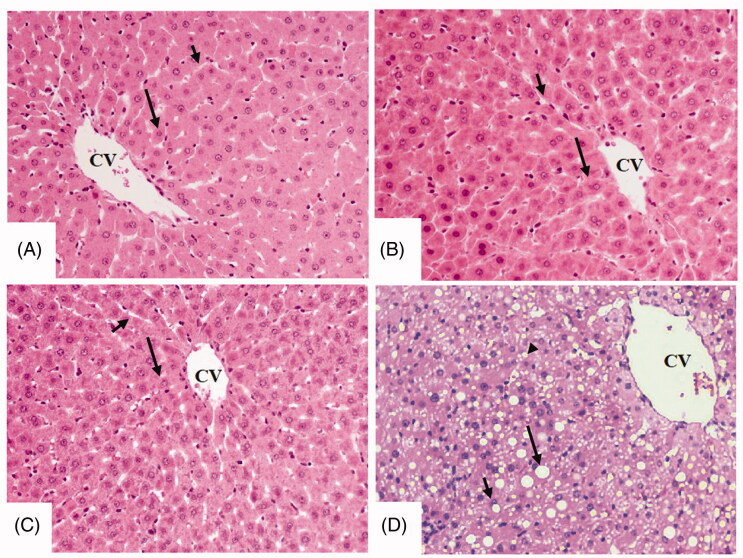
Figure 1.
Histological sections from some experimental groups of rats as stained by haematoxylin and eosin (H &E). A, B, and C were taken from control, control + ellagic acid (EA), and control + EA + compound C (CC/AMPK inhibitor), respectively and showing normal liver architectures with rounded normally-appeared hepatocytes (long arrow) radiating from the central vein (CV) with intact sinusoids (short arrow). D was taken from an STZ-diabetic rat (T1DM-induced rat) and showed severe cytoplasmic accumulation of lipid vacuoles of different sizes including large (long arrow), medium (short arrow), and small (arrowhead) vacuoles. 200×.

