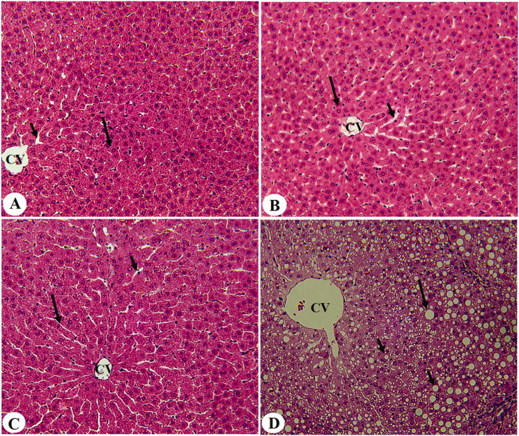
Figure 2.
Histological sections from some experimental groups of rats as stained by haematoxylin and eosin (H &E). A was taken for T1DM-induced rats and showed severe congestion in the portal vein (PV) that is surrounded by increased infiltration of inflammatory cells (curved arrow). B was taken from a T1DM + EA-treated rat and showed almost normal architectures like those seen in the control rats with normal hepatocytes (long arrow). However, very few cytoplasmic lipid accumulations are still seen in few hepatocytes. C and D were taken from T1DM + EA + CC and showed similar abnormal features like those observed in T1DM-induced rats including increased accumulation of large, medium, and small cytoplasmic lipid droplets (long arrow, short arrow, and arrowheads, respectively), with increased accumulation of inflammatory cells around the congestive portal vein (PV) (curved arrow).

