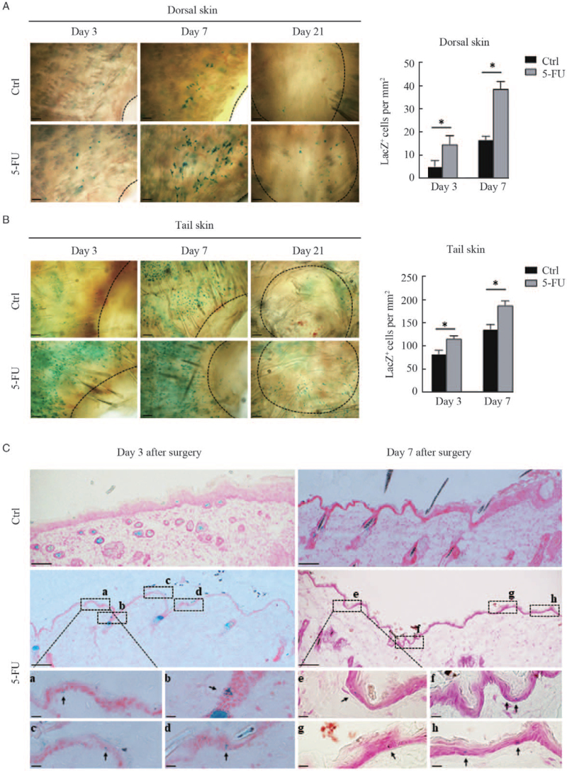
Figure 1.
Visualization of LacZ+ cells in the margins of wounds treated with or without 5-FU. Full-thickness excisional wounds were created in the dorsal and tail skins of Dct-lacZ transgenic mice. The skin was biopsied from the wound margins on the third, seventh, and 21st day after surgery. Representative images of LacZ+ cells in dorsal skin (A, upper panel) and tail skin (B, lower panel) by whole-mount X-Gal staining are shown. The black dashed lines indicate the leading edge of wounds. Scale bars: 100 μm. Histograms showing the number of LacZ+ melanocytes in the wound margins of dorsal and tail skin on the third and seventh day after surgery, expressed as means ± SD of three independent experiments, ∗P < 0.01, compared to controls. (C) Representative images of LacZ+ cells in cryosections of dorsal skin with X-Gal staining are shown. Scale bars: 100 μm. Black rectangles (a–h) indicate regions enlarged below. Scale bar: 20 μm (enlarged images). Black arrows indicate typical LacZ+ melanocytes. 5-FU: 5-fluorouracil; SD: Standard deviation.

