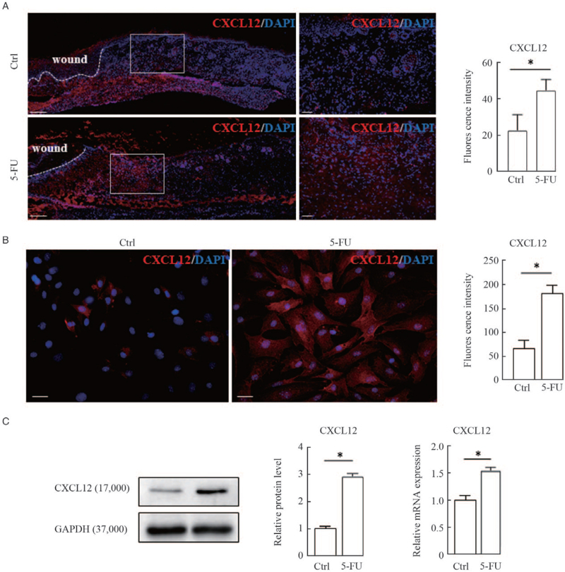
Figure 2.
Upregulation of chemokine CXCL12 expression in wound margins and cultured fibroblasts by 5-FU. (A) Representative images of immunofluorescence staining of CXCL12 (red) and counterstaining with DAPI (blue) in cryosections of dorsal skin wounds are shown. White dashed lines indicate the leading edge of wounds. Scale bars: 200 μm. White rectangles indicate regions enlarged on the right. Scale bar: 50 μm (enlarged images). Histograms showing the comparison of fluorescence intensity for wound margins treated with or without 5-FU are shown on the right. Immunofluorescence staining (B), Western blotting and qRT-PCR (C) were used to examine the expression levels of CXCL12 protein and mRNA in cultured fibroblasts treated with 50 μmol/L 5-FU for 24 h. Representative blots are shown. Data are shown as means ± SD of three independent experiments. ∗P < 0.01 vs. control. 5-FU: 5-fluorouracil; DAPI: diamidino-2-phenylindole; qRT-PCR: Quantitative real-time polymerase chain reaction; SD: Standard deviation.

