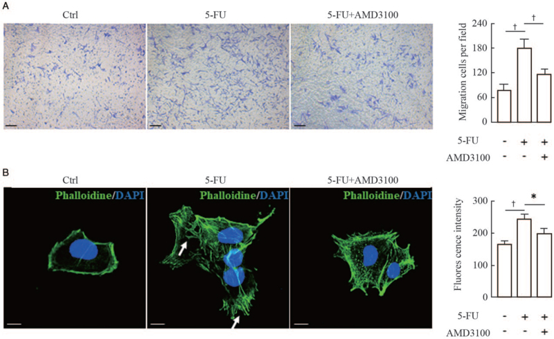
Figure 4.
Effect of AMD3100 (a specific CXCR4 antagonist) on the migration and F-actin distribution of melanocytes. (A) Melan-a melanocytes were seeded into the upper chambers and pretreated with 10 μmol/L AMD3100 for 1 h, and the lower chambers were seeded with murine fibroblasts for 24 h of coculture, as described in the Materials and Methods Section. Cells that migrated to the lower surface of the inserts were counted in five microscopic fields using a 20 × objective in at least three independent experiments. Representative micrographs of migrating cells are shown. Scale bars: 50 μm. Histogram (on the right) shows the number of migrated cells in five random microscopical fields. (B) Melan-a melanocytes growing on glass coverslips were treated with or without 10 μmol/L AMD3100 for 1 h, and were then treated with the conditioned medium from 5-FU-treated fibroblasts and subsequently stained for actin with FITC-phalloidin (green). Nuclei were counterstained with DAPI (blue). Representative images of F-actin staining are shown. White arrows indicate the distribution of coarse actin bundles in the cytoplasm. Scale bars: 10 μm. Histogram (on the right) shows the fluorescent intensities of 20 cells. Data represent means ± SD of three independent experiments. ∗P < 0.05 and †P < 0.01 vs. 5-FU group. 5-FU: 5-fluorouracil; DAPI: Diamidino-2-phenylindole; F-actin: Filamentous actin; SD: Standard deviation.

