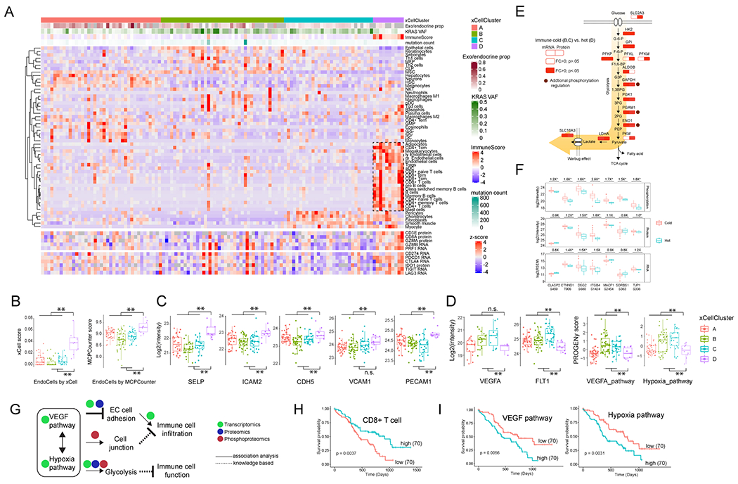
Figure 6. Delineation of the cellular composition of PDAC tumors and identification of biological events accounting for the immune-cold phenotype.

A) The 140 tumors were classified into four clusters based on tumor composition (upper heatmap). The cytotoxic T cells, together with endothelial cells enriched in the cluster D are highlighted by a rectangle. The expression of immune cytotoxic factor and checkpoint genes is shown in the sample order (lower heatmap). B) The comparison of endothelial cells between immune hot and cold samples based on the in silico deconvolution using either xCell or MCPCounter. C) Immune cold tumors have reduced endothelial adhesion proteins. D) Immune cold tumors have upregulated VEGF and hypoxia pathways. B-D: **p<0.01, n.s. not significant, Student’s t-test. E) The immune cold tumors had higher levels of glycolytic pathway components. Shown are the comparison of these components between immune cold vs hot at both the RNA and protein level. Some of the pathway components are identified with known functional phosphosites and are highlighted by brown circles. F) Phosphorylation pathway enrichment showed that the immune cold samples have higher phosphorylation levels of cell junction proteins. Shown are immune cold vs. hot fold changes for protein phosphorylation, protein expression and RNA expression. *p<0.05, Student’s t-test. G) The possible working model. VEGF and hypoxia pathways are associated with aberrant tumor vasculature and a hypoxic tumor microenvironment, and downregulated endothelial cell adhesion proteins, increased glycolysis and cell junction further inhibit the cytotoxic immune infiltration and function. H) The clinical outcome associated with CD8+ T cells. I) The clinical outcome associated with VEGF and hypoxia pathway activities. H-I): The p values were derived from logrank test and numbers in parentheses represent sample sizes for each group.
