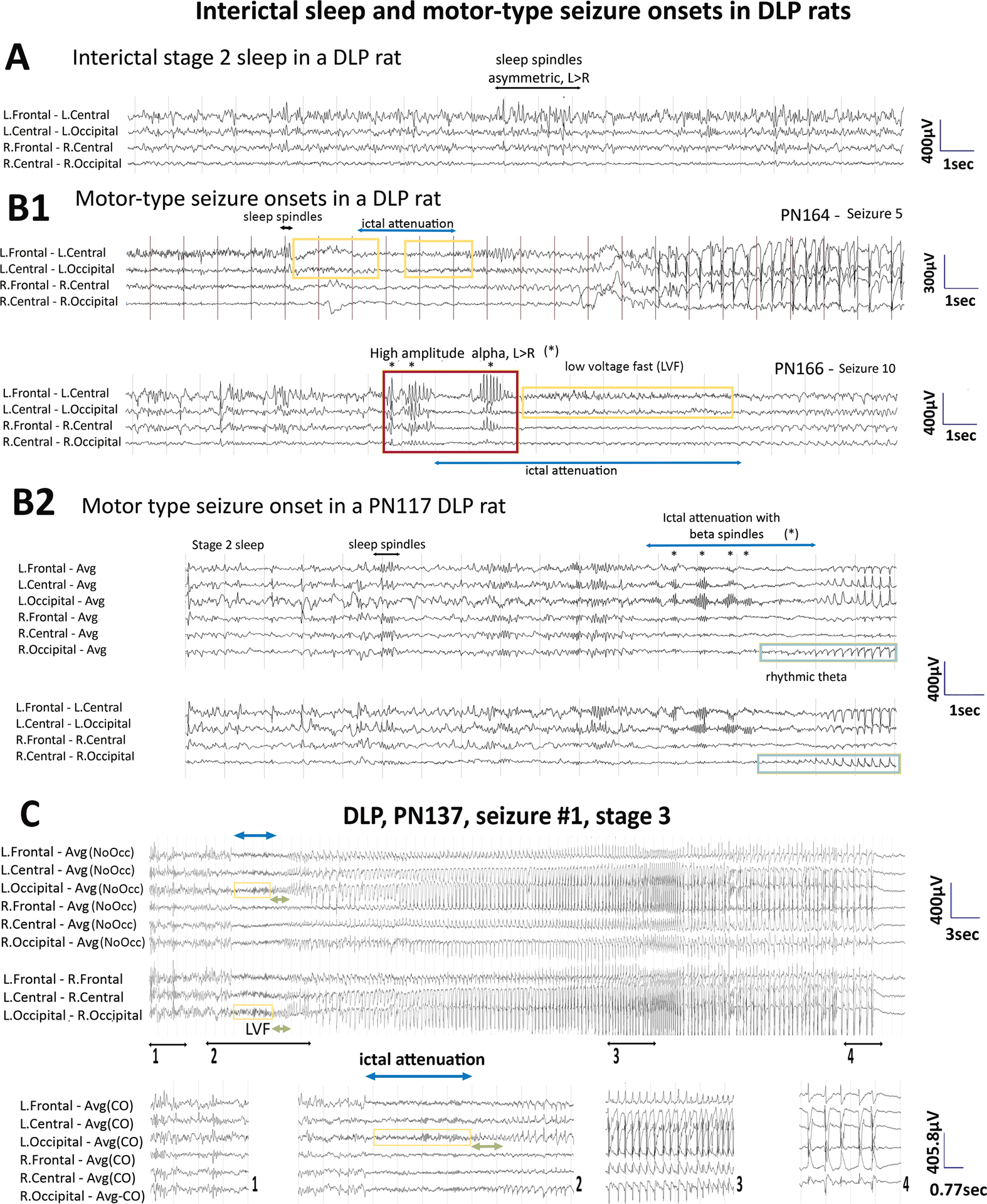
Fig 2. Electrographic patterns during interictal slow wave sleep (SWS) (A), motor-type seizure onsets (B) and motor-type seizure patterns (C) in DLP adult rats.

Panel A: Interictal SWS EEG of a PN164 DLP rat shows asymmetric background with more prominent sleep spindles at the left frontocentral region (black horizontal arrow).
Panel B: Onsets of motor-type seizures captured in two DLP rats. (B1) Seizure onsets from PN164 and PN166 are from the rat from panel A. Asymmetric sleep spindles in preictal SWS (black horizontal arrow) precede seizure onset in seizure 5 (PN164). The yellow boxes indicate the low voltage fast (LVF) at the onsets of seizure 5 and 10 and the cyan lines indicated ictal attenuations. Seizure 10 of the same rat starts with high amplitude alpha bursts (*, red box) with higher amplitude and sharper contour compared to the usual sleep spindles, more prominent at the left than the right hemisphere, maximal frontally. (B2) Seizure onset of a PN117 DLP rat shows pre-ictal sleep spindles in SWS. Ictal attenuation (cyan line) and left occipital beta spindles (*) emerge followed by rhythmic theta (green boxes) at the right occipital region that evolves into the seizure pattern.
Panel C: EEG of the first detected motor-type seizure (stage 3) of a PN137 adult DLP rat. The pre-ictal EEG shows asymmetric sleep spindle (maximal left frontocentral) (segment 1). Ictal attenuation at seizure onset (segment 2) with left occipital LVF leading into rhythmic theta which spreads to bilateral occipital regions evolving into a seizure pattern bilaterally (segment 3) which terminates with postictal attenuation (segment 4). EEG is presented with either average montage excluding occipital leads or transverse montage.
Sampling rate: 200Hz. Low frequency filter (LFF): 1Hz, high frequency filter (HFF): 70Hz. Avg: Average reference; Avg (NoOcc): average reference with occipital leads excluded; L: left, R: right.
