
Fig 4. Sleep-wake periods and histological brain findings in adult rats.
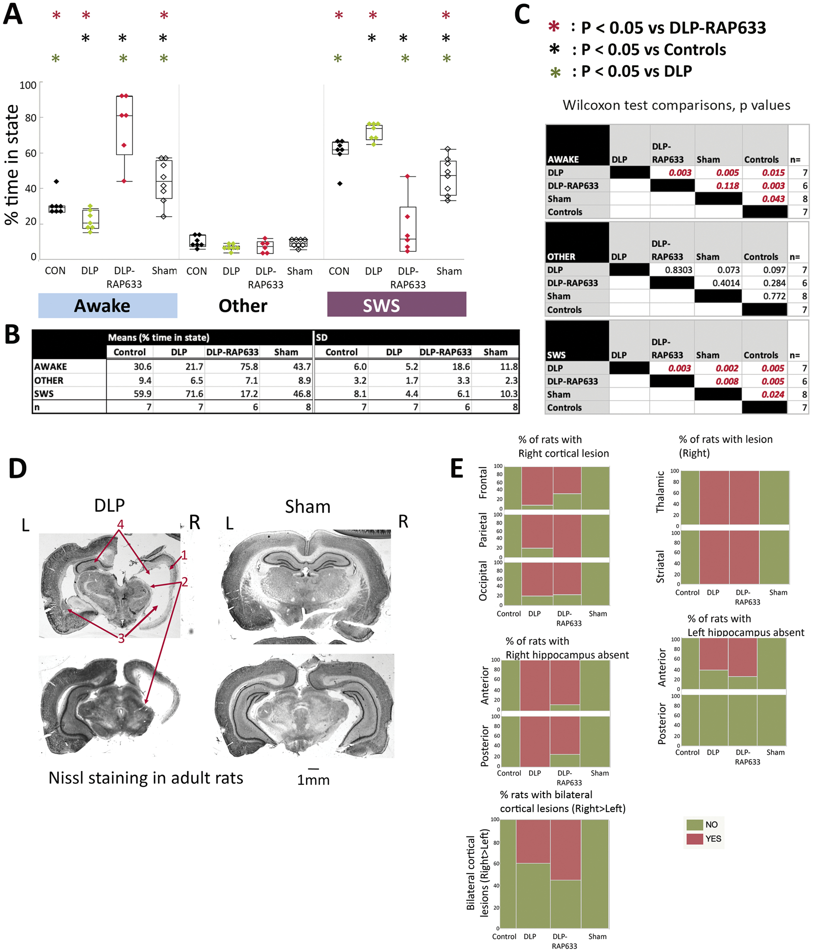
Panels A-C: Percentage of slow wave sleep (SWS) and awake state periods in DLP, DLP-RAP633, control, and sham rats during the light periods, aged PN135–177. REM and passive wakefulness were scored as “other” due to the lack of electromyographic recordings. Panels A and B present the means and standard deviations (SD) of % time spent in each state for each group. Panel C shows the nonparametric Wilcoxon test comparisons across treatment groups for each state (distributions were not normal). All studied treatment groups (DLP, DLP-RAP633, sham) were different than controls in the SWS and awake states. DLP rats had increased SWS and reduced awake states from every other group. In contrast, DLP-RAP633 had significantly increased time spent in wakefulness compared to every other group. Sham rats spent more time in awake and less in SWS state compared to either controls or DLP groups but had shorter awake periods compared to DLP-RAP633. DLP (n=7 rats); DLP-RAP633 (n=6 rats); control (n=7 rats); sham (n=8 rats).
Panel D: Nissl staining of coronal sections of adult rat brains in the DLP and sham groups. The DLP brain shows lesion at the right cortical, thalamic and striatal regions, absence of right and atrophy of left hippocampus. L: Left; R: Right. The barscale corresponds to 1mm distance. The red arrows indicate the right cerebral cortex (1), right thalamic regions (2), left striatum and corresponding region at the right (3), left hippocampus and corresponding region at the right (4).
Panel E: The distribution of injury / atrophy in the brains per each group is shown here for cortical, hippocampal, and thalamic / striatal regions or bilateral cortical lesions. Injury is classified as present (“yes”) or absent (“no”). The injury was consistently seen at the right cortical, hippocampal and striatal / thalamic periventricular regions in the DLP and DLP-RAP633 rats, unlike control and sham rats that had no injury. Detailed statistical comparisons are given in the Supplementary Table 2.