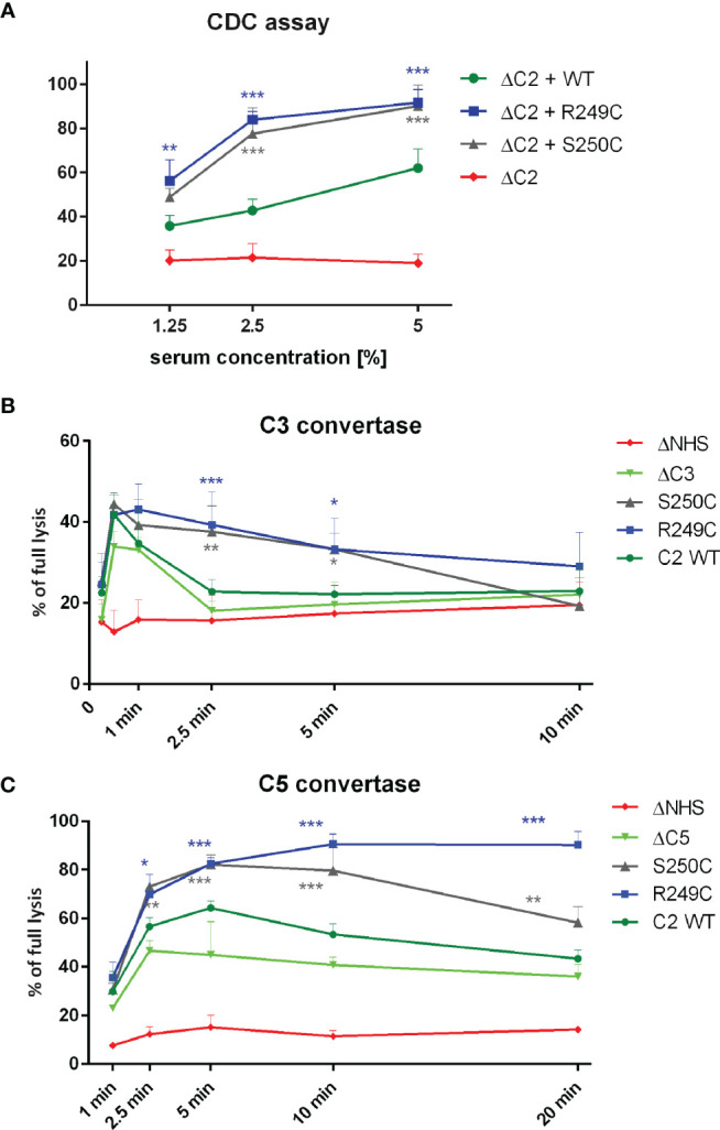
Figure 1.

Functional assays of C2 variants. (A) CDC assay. Calcein-loaded Raji cells sensitized with anti-CD20 mAb (ofatumumab) were suspended in C2-depleted serum (ΔC2) supplemented with physiological concentration of C2 protein. Cells were incubated for 30 min at 37°C. Supernatant was collected, and fluorescence of released calcein was measured. The readout obtained for cells lysed with 30% DMSO was considered as full lysis, and the readout obtained for cells incubated with ΔC2 serum alone indicated background lysis (negative control). (B, C) Convertase activity assays for classical C3 convertase (B) and classical C5 convertase (C) were performed on calcein-loaded Ramos cells suspended in 15% of C3-depleted serum (ΔC3) (B) or C5-depleted serum (ΔC5) (C) supplemented with physiological concentration of C2 variants. After indicated time period, cells were washed with EDTA-containing buffer to disable further convertase formation and then suspended in 5% guinea pig serum diluted in the same EDTA buffer. The readout was performed as in CDC assay, but heat-inactivated serum (ΔNHS) instead of ΔC2 served as a negative control. Data are collected from at least three experiments. Symbols *, **, and *** denote statistically significant differences vs. WT supplementation, at p levels of 0.05, 0.01, and 0.001 according to Dunn’s multiple comparison test.
