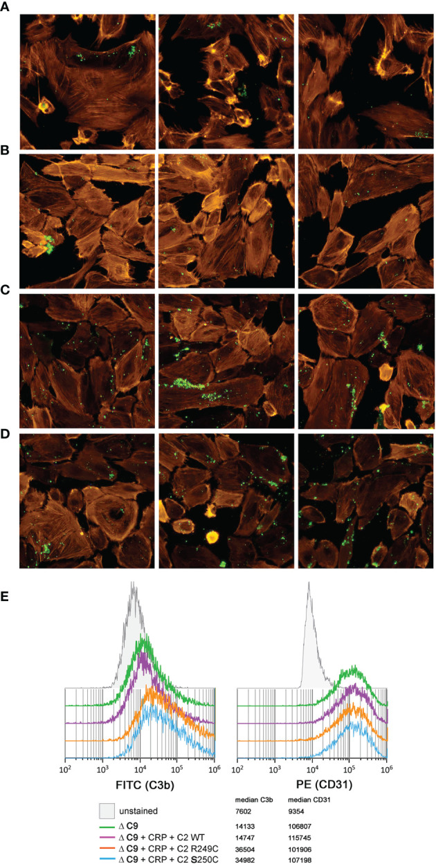
Figure 3.

C3b deposition on glomerular endothelial cells. Immortalized glomerular endothelial cells were seeded onto glass chambers and exposed to C9-depleted human serum (ΔC9) in order to prevent lysis. Deposition of C3b (green) was analyzed by fluorescent microscopy (A–D) when nothing (A), CRP + WT C2 (B), CRP + R249C variant (C), or CRP + S250C variant (D) was added. Labeled phalloidin (orange) was used as a counterstaining. Three independent images are shown for each condition. (E) To perform an alternative, quantitative analysis that confirms the observed pattern of immunofluorescence, cells were harvested and examined by flow cytometry for both C3b deposition (left histogram) and CD31 expression (right histogram). Results show 10,000 events, previously gated to eliminate cell doublets and cell debris.
