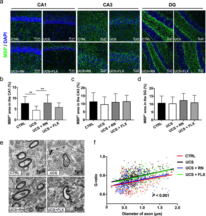
Fig. 6. The effects of running exercise and fluoxetine on the myelination in the hippocampus.
a Immunohistochemical staining for MBP+ (green) myelinated fibers in the CA1, CA3, and DG subfields of the hippocampus. DAPI (blue) was used as a nuclear counterstain. Scale bar = 50 μm. b–d Immunofluorescence analyses of MBP+ (green) myelinated fibers in the CA1, CA3, and DG hippocampal subfields of mice in the CTRL group, UCS group, UCS + RN group, and UCS + FLX group (n = 5 for each group). e Electron micrographs of axons in the CA1 hippocampal subfield of mice in the CTRL group, UCS group, UCS + RN group, and UCS + FLX group. Scale bar = 1 μm. f Quantification of the myelin sheath thickness (G-ratio) in the CA1 subfield of mice in the CTRL group (n = 303 axons from four mice), UCS group (n = 316 axons from four mice), UCS + RN group (n = 332 axons from four mice), and UCS + FLX group (n = 310 axons from four mice). All data are shown as the means ± SDs. * P < 0.05 and ** P < 0.01.

