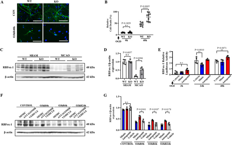
Fig. 4. CRTC1 deletion and miR-132 knockdown aggravate neuron damage.
A Representative microtubule-associated protein 2 (MAP2) immunostaining images of CRTC1 KO and WT neurons, before and after OGD. (Scale Bar, 100 μm). B LDH assay of CRTC1 KO and WT neuron up to 48 h after ischemia/reperfusion. Representative Western Blot images (C) and quantification (D) of RBFox-1 in CRTC1 KO and WT mice penumbra region. E Relative luciferase activity of CHO cell line co-transfected with RBFox-1 and miR-132 vehicle/mimic/antagomir. Representative Western Blot images (F) and quantification (G) of RBFox-1 in neurons transfected with miR-132 mimic/antagomir/vehicle, before and after OGD.

