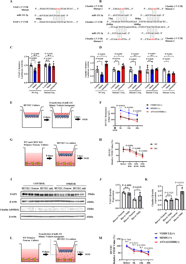
Fig. 7. Neuron secretes miR-132/212 to endothelial cells to alleviate BBB breakdown after ischemia/reperfusion.
The partial sequence of miR-132 and its one putative binding site in the 3′-UTR of the mouse TJAP-1 (A) and four putative binding sites in the 3′-UTR of the mouse Claudin-1 (B), with each mutant 3′-UTR. Quantitative data showed that transfection of miR-132 mimic decreased luciferase activity of the reporter vector containing wild-type 3′-UTR sequence of mouse TJAP-1 (C) and Claudin-1 (D), while transfection of miR-132 mimic or antagomir had no effects on the luciferase activity of the reporter vector containing mutation of 468-470 bp within mouse TJAP-1 3′-UTR (C) and mutation of 1844-1846 bp within mouse Claudin-1 3′-UTR (D). (E) HUVECs were transfected with miR-132 mimic/vehicle/antagomir, then subjected to OGD. (F) TEER evaluation indicates that miR-132 mimic significantly attenuated the decline in TEER values after OGD, while miR-132 antagomir aggravated HUVEC resistance to hypoxia. n = 3 for each group. (G) A BBB model in vitro was constructed by a co-culture system of HUVECs and neurons derived from CRTC1 KO and WT mice. (H) HUVECs co-cultured with CRTC1 KO neurons exhibited progressively decreased TEER values after OGD compared with HUVECs co-cultured with WT neurons. Representative Western Blot images (I) and quantification of TJAP-1 (J) and Claudin-1 (K) of HUVECs co-cultured with or without neurons, before and after OGD. (L) HUVECs were co-cultured with neurons that were transfected with miR-132 mimic/antagomir/vehicle, then subjected to OGD. (M) TEER evaluation indicates that miR-132 mimic by paracrine secretion significantly attenuated the decline in ECs TEER values after OGD. n = 10 for each group.

